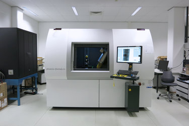

Inside X-ray tomography machine
Peering through the protective lead glass window of ESA's X-ray tomography machine, where a 3D-printed mirror is being X-rayed.
The machine is the engineering equivalent of a medical CT scanner – and does an equivalent job, revealing the details of the interior of a test item without destroying it in the process.
The test part is slowly moved around as a thousand X-ray images are taken. Then specialised software stacks these individual images into a detailed 3D model.
Total acquisition time depends on the size of the part – it might be completed in six to seven hours. For a larger part multiple acquisitions might need to be performed across different sections of the part, then merged.
The resulting 3D model has a spatial resolution down to around 0.03 mm, portraying not only its surface but also its interior features, so that the engineers can perform a detailed flythrough around – and deep inside – the test part.


